Home

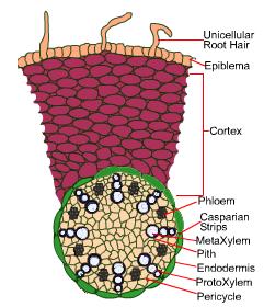
Bezchybný vstúpiť Typ predku transverse section of a root schematic život skontrolovať vyžarovať

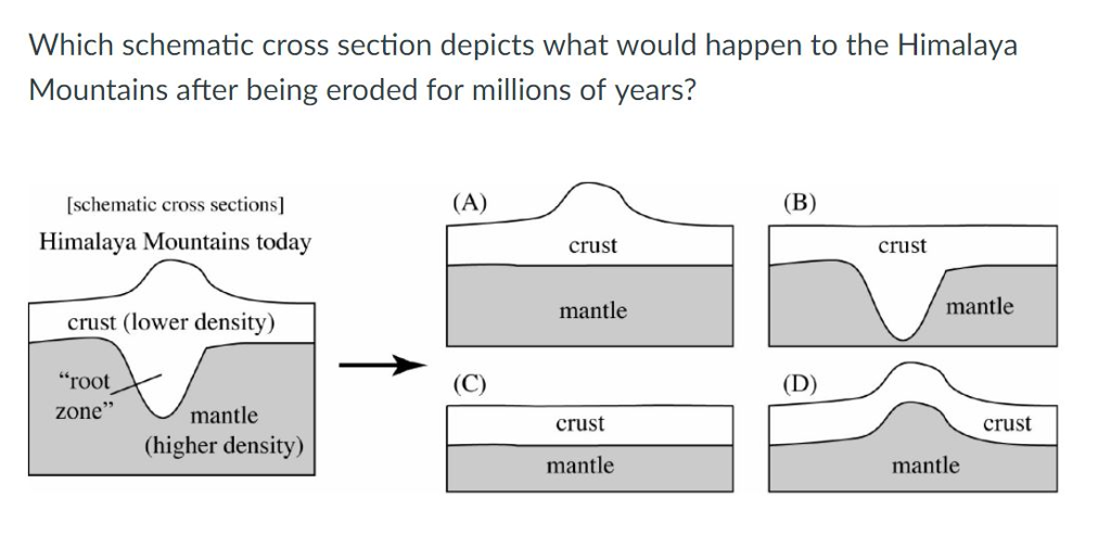
Solved Which schematic cross section depicts what would | Chegg.com

Draw a neat labelled diagram of Transverse Section of young 'Dicot Root'  and explain different parts. - Sarthaks eConnect | Largest Online Education  Community
Draw a neat labelled diagram of Transverse Section of young 'Dicot Root' and explain different parts. - Sarthaks eConnect | Largest Online Education Community
File:Root(cross section).jpg - Wikimedia Commons

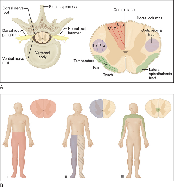
SPINAL DISEASE: NEOPLASTIC, DEGENERATIVE, AND INFECTIVE SPINAL CORD  DISEASES AND SPINAL CORD COMPRESSION | Neupsy Key
SPINAL DISEASE: NEOPLASTIC, DEGENERATIVE, AND INFECTIVE SPINAL CORD DISEASES AND SPINAL CORD COMPRESSION | Neupsy Key

Schematic Cross-Section of a Primary Root (upper bubble = transmembrane...  | Download Scientific Diagram
Schematic Cross-Section of a Primary Root (upper bubble = transmembrane... | Download Scientific Diagram
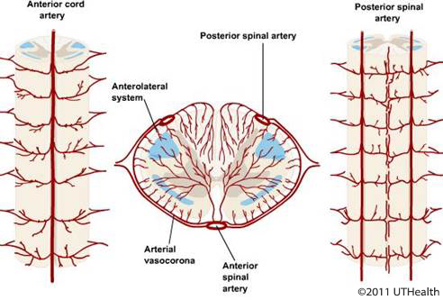
Neuroanatomy Online: An Open Access Electronic Laboratory for the  Neurosciences | Introduction - Overview of the Nervous System
Neuroanatomy Online: An Open Access Electronic Laboratory for the Neurosciences | Introduction - Overview of the Nervous System

Schematic drawing of root longitudinal section showing the different... |  Download Scientific Diagram
Schematic drawing of root longitudinal section showing the different... | Download Scientific Diagram

Botany | Life Science & Biomedical | Carlson Stock Art | Biology plants,  Botany, Plant science
Botany | Life Science & Biomedical | Carlson Stock Art | Biology plants, Botany, Plant science

Cross Section Through A Molar Tooth Showing The Crown And Root Plus The  Gum, Bone, Blood Vessels And Nerves. Stock Photo, Picture And Royalty Free  Image. Image 70263895.
Cross Section Through A Molar Tooth Showing The Crown And Root Plus The Gum, Bone, Blood Vessels And Nerves. Stock Photo, Picture And Royalty Free Image. Image 70263895.

Electron-microscopic structure of protozoa . Text-figure 15. Schematic  drawing showing the organization of adoral membranelles of Stentor. C  cilia; K kinetosomes; R root fibrils; Rbf, bundle of root fibrils from a
Electron-microscopic structure of protozoa . Text-figure 15. Schematic drawing showing the organization of adoral membranelles of Stentor. C cilia; K kinetosomes; R root fibrils; Rbf, bundle of root fibrils from a

Schematic cross-sections of a primary root (A) and a mature root with... |  Download Scientific Diagram
Schematic cross-sections of a primary root (A) and a mature root with... | Download Scientific Diagram

Schematic cross-section of a root. Similarly shaped and shaded cells... |  Download Scientific Diagram
Schematic cross-section of a root. Similarly shaped and shaded cells... | Download Scientific Diagram
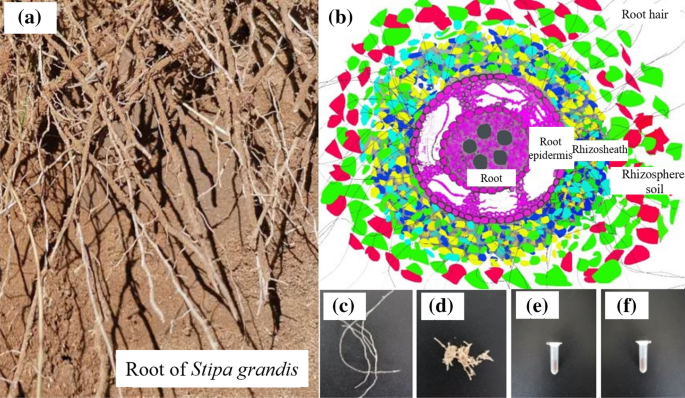
Isolation of rhizosheath and analysis of microbial community structure  around roots of Stipa grandis | Scientific Reports
Isolation of rhizosheath and analysis of microbial community structure around roots of Stipa grandis | Scientific Reports

Monocot Root: A Complete Anatomy with Diagram

View Image
View Image

Schematic representation of root sections in almonds roots. Schematic... |  Download Scientific Diagram
Schematic representation of root sections in almonds roots. Schematic... | Download Scientific Diagram

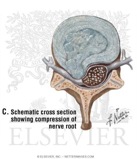
Schematic Cross Section Showing Compression of Nerve Root

Schematic diagrams of tissues of seedling root and growing young... |  Download Scientific Diagram
Schematic diagrams of tissues of seedling root and growing young... | Download Scientific Diagram

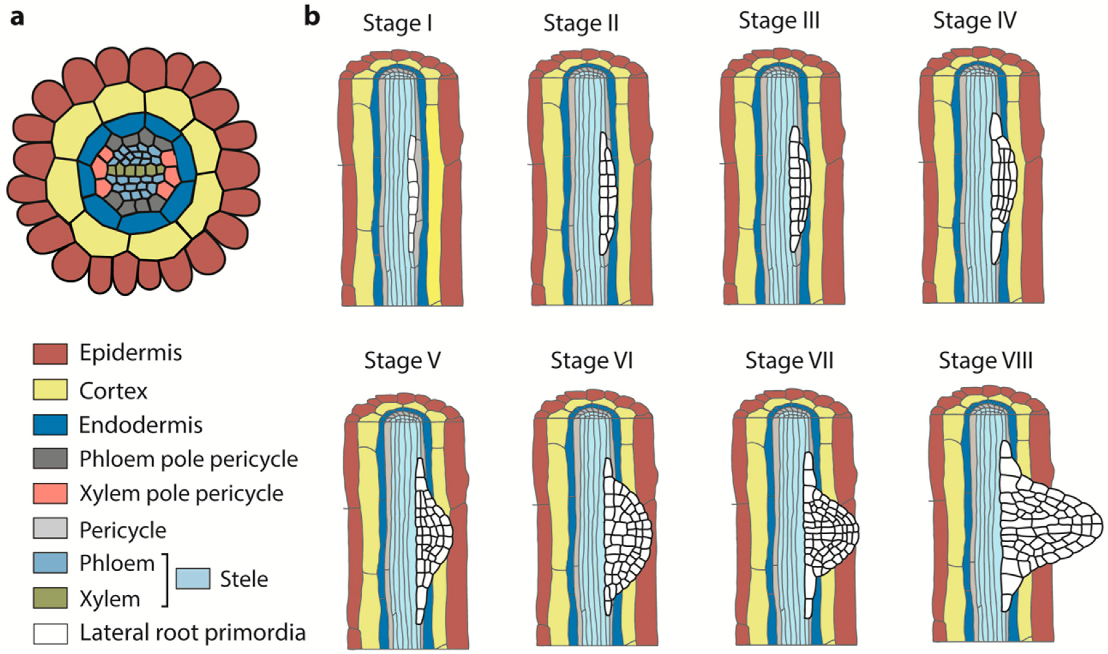
IJMS | Free Full-Text | Interplay of Auxin and Cytokinin in Lateral Root  Development
IJMS | Free Full-Text | Interplay of Auxin and Cytokinin in Lateral Root Development

Parsimonious Model of Vascular Patterning Links Transverse Hormone Fluxes  to Lateral Root Initiation: Auxin Leads the Way, while Cytokinin Levels Out  | PLOS Computational Biology
Parsimonious Model of Vascular Patterning Links Transverse Hormone Fluxes to Lateral Root Initiation: Auxin Leads the Way, while Cytokinin Levels Out | PLOS Computational Biology

Pin on Les Racines
Pin on Les Racines

Deciphering the genetic basis of wheat seminal root anatomy uncovers  ancestral axial conductance alleles | bioRxiv
Deciphering the genetic basis of wheat seminal root anatomy uncovers ancestral axial conductance alleles | bioRxiv
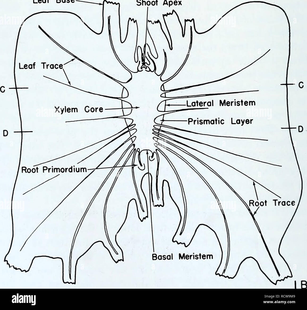
The developmental anatomy of Isoetes. Isoetes; Botany. ateral Meristem  Prismatic Layer Basal Meristem Root Primordium Leaf Base Shoot Apex. Fig.  1. The principal planes of sectioning for two-lobed plants. A. Furrow
The developmental anatomy of Isoetes. Isoetes; Botany. ateral Meristem Prismatic Layer Basal Meristem Root Primordium Leaf Base Shoot Apex. Fig. 1. The principal planes of sectioning for two-lobed plants. A. Furrow
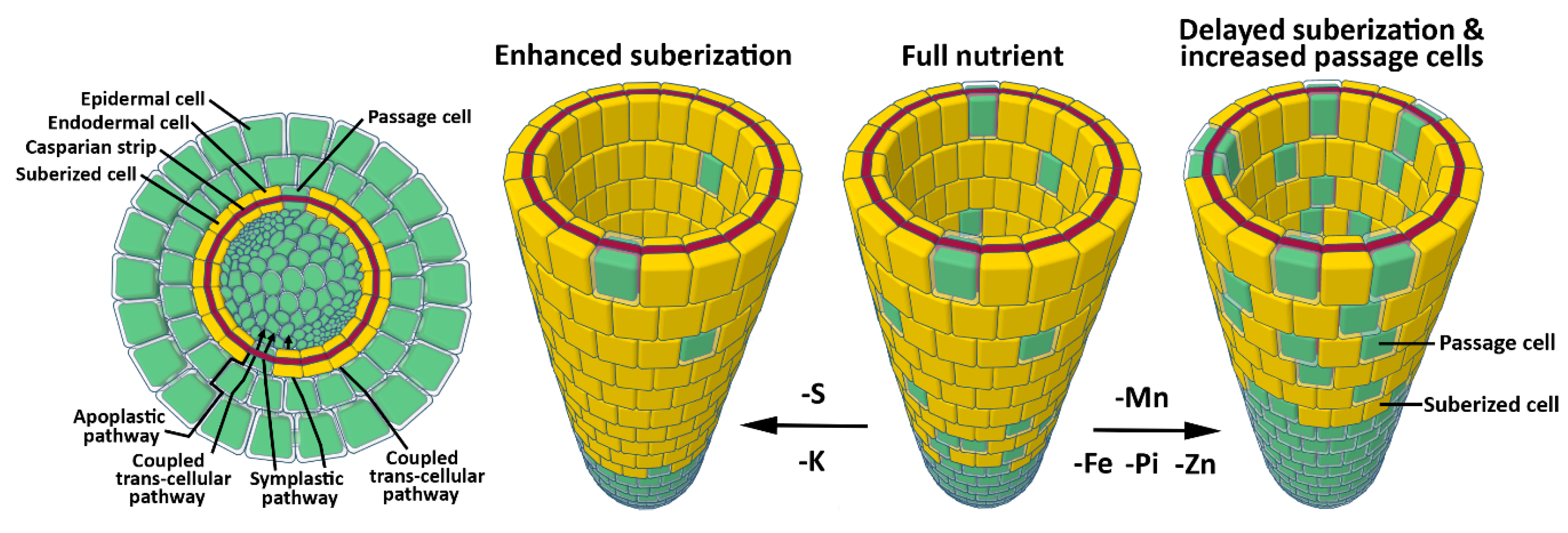
Cross section of a young root (Modified from Fahn 1987). | Download  Scientific Diagram
Cross section of a young root (Modified from Fahn 1987). | Download Scientific Diagram

IJMS | Free Full-Text | Feeding the Walls: How Does Nutrient Availability  Regulate Cell Wall Composition?
IJMS | Free Full-Text | Feeding the Walls: How Does Nutrient Availability Regulate Cell Wall Composition?

An atlas of the differential diagnosis of the diseases of the nervous  system; analytical and semeiological neurological charts . I n. L9 Schematic  representation of brain stem; showing nuclei and nerve
An atlas of the differential diagnosis of the diseases of the nervous system; analytical and semeiological neurological charts . I n. L9 Schematic representation of brain stem; showing nuclei and nerve